How to Prepare

Before scheduling a mammogram, the American Cancer Society (ACS) and other specialty organizations recommend that you discuss any new findings or problems in your breasts with your doctor. In addition, inform your doctor of any prior surgeries, hormone use, and family or personal history of breast cancer.

Do not schedule your mammogram for the week before your period if your breasts are usually tender during this time. The best time for a mammogram is one week following your period. Always inform your doctor or x-ray technologist if there is any possibility that you are pregnant.

The ACS also recommends you:

  • Do not wear deodorant, talcum powder or lotion under your arms or on your breasts on the day of the exam. These can appear on the mammogram as calcium spots.
  • Describe any breast symptoms or problems to the technologist performing the exam.
  • If possible, obtain prior mammograms and make them available to the radiologist at the time of the current exam.
  • Ask when your results will be available; do not assume the results are normal if you do not hear from your doctor or the mammography facility.

3D MammographyUnited Radiology Coach

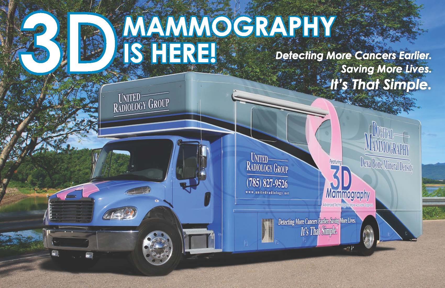
United Radiology Group has recently increased their services at TCLMH and other rural Kansas hospitals with the purchase of a new 3D mobile mammography coach. 3D mammography provides better quality exams, earlier breast cancer detection, and reduces unnecessary call backs by up to 40%. This new technology allows doctors to examine breast tissue layer by layer instead of a flat 2D image and detects 41% more invasive breast cancers. Women of all ages and breast densities benefit from this new service.

The new 3D Mammography coach features:

• Patient entrance
• Waiting area
• Reception/check-in desk
• Private changing rooms
• Wheelchair access door
• Private screening room

The mammography coach provides services once a month at TCLMH. Although most insurance plans cover the cost of 3D mammography, it’s important to check with your insurance provider first before scheduling. To obtain a 3D mammogram, please call the radiology department at (785)743-3411.

Patients must alert the technologist if they may be pregnant!


How is the procedure performed?

Mammography is performed on an outpatient basis.

During mammography, a specially qualified radiologic technologist will position your breast in the mammography unit. Your breast will be placed on a special platform and compressed with a paddle (often made of clear Plexiglas or other plastic). The technologist will gradually compress your breast.

Breast compression is necessary in order to:

  • Even out the breast thickness so that all of the tissue can be visualized.
  • Spread out the tissue so that small abnormalities are less likely to be obscured by overlying breast tissue.
  • Allow the use of a lower x-ray dose since a thinner amount of breast tissue is being imaged.
  • Hold the breast still in order to minimize blurring of the image caused by motion.
  • Reduce x-ray scatter to increase sharpness of picture.

You will be asked to change positions between images. The routine views are a top-to-bottom view and an angled side view. The process will be repeated for the other breast.


How Does It Work?

X-rays are a form of radiation like light or radio waves. X-rays pass through most objects, including the body. Once it is carefully aimed at the part of the body being examined, an x-ray machine produces a small burst of radiation that passes through the body, recording an image on photographic film or a special detector.

Different parts of the body absorb the x-rays in varying degrees. Dense bone absorbs much of the radiation while soft tissue, such as muscle, fat and organs, allow more of the x-rays to pass through them. As a result, bones appear white on the x-ray, soft tissue shows up in shades of gray and air appears black.

Until recently, x-ray images were maintained as hard film copy (much like a photographic negative). Today, most images are digital files that are stored electronically. These stored images are easily accessible and are frequently compared to current x-ray images for diagnosis and disease management.