How to Prepare

You may be asked to wear a gown during the exam or you may be allowed to wear your own clothing.

Women should always inform their physician or technologist if there is any possibility that they are pregnant or if they are breastfeeding. See the Safety page for more information about pregnancy and breastfeeding related to nuclear medicine imaging.

You should inform your physician and the technologist performing your exam of any medications you are taking, including vitamins and herbal supplements. You should also inform them if you have any allergies and about recent illnesses or other medical conditions.

Jewelry and other metallic accessories should be left at home if possible, or removed prior to the exam because they may interfere with the procedure.

You will receive specific instructions based on the type of scan you are undergoing.

In some instances, certain medications or procedures may interfere with the examination ordered.

Outsourced Services

Nuclear Medicine is provided by Front Range Nuclear Services every Monday.

Nuclear Medicine

Nuclear medicine is a branch of medical imaging that uses small amounts of radioactive material to diagnose and determine the severity of or treat a variety of diseases, including many types of cancers, heart disease, gastrointestinal, endocrine, neurological disorders and other abnormalities within the body. Because nuclear medicine procedures are able to pinpoint molecular activity within the body, they offer the potential to identify disease in its earliest stages as well as a patient's immediate response to therapeutic interventions.

100 0281

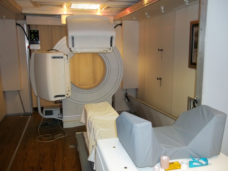
Nuclear medicine imaging procedures are noninvasive and, with the exception of intravenous injections, are usually painless medical tests that help physicians diagnose and evaluate medical conditions. These imaging scans use radioactive materials called radiopharmaceuticals or radiotracers.

Depending on the type of nuclear medicine exam, the radiotracer is either injected into the body, swallowed or inhaled as a gas and eventually accumulates in the organ or area of the body being examined. Radioactive emissions from the radiotracer are detected by a special camera or imaging device that produces pictures and provides molecular information.

In many centers, nuclear medicine images can be superimposed with computed tomography (CT) or magnetic resonance imaging (MRI) to produce special views, a practice known as image fusion or co-registration. These views allow the information from two different exams to be correlated and interpreted on one image, leading to more precise information and accurate diagnoses. In addition, manufacturers are now making single photon emission computed tomography/computed tomography (SPECT/CT) and positron emission tomography/computed tomography (PET/CT) units that are able to perform both imaging exams at the same time. An emerging imaging technology, but not readily available at this time is PET/MRI.

How Does It Work?

With ordinary x-ray examinations, an image is made by passing x-rays through the patient's body. In contrast, nuclear medicine procedures use a radioactive material, called a radiopharmaceutical or radiotracer, which is injected into the bloodstream, swallowed or inhaled as a gas. This radioactive material accumulates in the organ or area of your body being examined, where it gives off a small amount of energy in the form of gamma rays. Special cameras detect this energy, and with the help of a computer, create pictures offering details on both the structure and function of organs and tissues in your body.

Unlike other imaging techniques, nuclear medicine imaging exams focus on depicting physiologic processes within the body, such as rates of metabolism or levels of various other chemical activity, instead of showing anatomy and structure. Areas of greater intensity, called "hot spots," indicate where large amounts of the radiotracer have accumulated and where there is a high level of chemical or metabolic activity. Less intense areas, or "cold spots," indicate a smaller concentration of radiotracer and less chemical activity.